Nutritional Diagnostics
Step 0: Husbandry
|
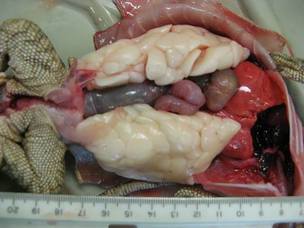
Poor husbandry has caused this savannah monitor to accumulate dangerous levels of visceral fat. Excess fat in healthy animals should be stored in the tail. (Courtesy savannahmonitor.net)
|
Step 2: Evaluate the DietEnsure that the animal is being fed a proper diet with proper supplementation. In order to do this, refer to the "species guide" section of this website.
|
Step 3: SymptomsUse the guide below to look at symptoms and what diseases they could be. There are often many possible results.
|
|
|
Symptoms:
Anorexia (refusal to eat) Obesity (make sure this is not ascites, pregnancy, a gravid animal or tumors) Deformaties/poor growth Lameness Muscle Tremors Bone Fractures Poor growth Eye infection/blindness Rash Sloughing skin Unable to apprehend food with tongue (frogs, salamanders) Bradycardia (slow heart rate) Swolen nodules under skin Goiter Seizure Flacid Paralysis Anemia |
Possible Diseases/Causes:
impaction, foreign body, poor husbandry, gastroenteritis, parasitic infection, obesity, fatty liver disease, hypovitaminosis A, vitamin E deficiency, renal failure, hypERvitaminosis A (amphibians) over feeding, improper diet, lack of burmation cycle, metabolic bone disease, rickets, protein deficiency, metabolic bone disease, rickets, gout metabolic bone disease, thiamin (B1) deficiency, vitamin E deficiency injury, metabolic bone disease, rickets protein deficiency, hypovitaminosis A hypovitaminosis A, thiamin (B1) deficiency, HYPERvitaminosis A, HYPERvitaminosis A, hypovitaminosis A (short tongue syndrome), thiamin (B1) deficiency, vitamin E deficiency, iodine deficiency and/or high goitrogen content in diet, iodine intoxication, vitamin E deficiency, vitamin E deficiency, hypERvitaminosis A, iron deficiency |
Nutritional Disease Information:
Metabolic Bone Disease:
Cause-Dietary imbalances of calcium, phosphorus and vitamin D. Can be caused by hypERvitaminosis A in amphibians.
Diagnostics-Physical examination, radiographs, low circulating calcium levels (extreme cases only)
Treatment-Provide proper lighting for species (often full spectrum UVB lighting) and provide exposure to direct sunlight for 30 minutes a day. Splint any broken bones. Increase dietary calcium while reducing dietary phosphorus. In anorexic patients, admister oral calcium gluconate. In severe cases a parietal vitamin D3 injection is recommended.
Rickets:
Cause-Lack of proper vitamin D supplementation through diet and/or UVB lighting.
Diagnostics-Physical examination, radiographs
Treatment-Although there is no calcium deficiency in the case of rickets, it should still be treated as metabolic bone disease as there is little ability to differentiate the diseases
Metastatic Mineralization:
Cause-A nutritional cause of this disease is not certain, but it is related to calcium metabolism. It was previously thought that this was a manifestation of hypervitaminosis D3, but this is no longer thought to be the case as little D3 is absorbed from the digestive tract of green iguanas, which commonly get metastatic mineralization. This disease is highly linked to renal failure.
Diagnostics-Radiographs show radiopacity of the aorta and possibly enlarged kidneys, blood work to confirm impaired renal function, serum calcium >40mg/dl in males and females not in egg development
Treatment-Salmon-calcitonin combined with diuresis with saline at 10 ml/kg every 24 hours. Ensure species appropriate UVB lighting and exposure to direct sunlight. Every effort to relieve the burden on the kidneys should be made by decreasing phosphorus and protein content of the diet.
Gout:
Cause-The exact cause in reptiles is not known but it is believed to be some combination of excess improper amino acids, low dietary potassium and/or high phosphorus. Renal failure is also implicated in gout.
Diagnostics-physical exam for swelling of joints, radiographs to see enlarged kidneys and radiodense material around joints, high serum uric acid, changes in plasma calcium and phosphorus, aspiration of swollen joint to visualize urates using microscopy
Treatment-increase hydration through husbandry, allopurinol may be helpful, increase dietary potassium and decrease protein content of the diet.
Renal Failure:
Cause-Numerous, but can be triggered by a high phosphorus diet and dehydration. High protein is not typically indicated, but should still be a consideration in herbivorous and omnivorous species.
Diagnostics-Blood work, low plasma calcium, elevated phosphorus and potassium, possibly elevated uric acid, certain, AST, ALT and CPK. Further diagnostics, such as radiographs, to determine the type of renal failure may be warranted.
Treatment-Ensure the animal is hydrated without over hydrating. Calcium gluconate should be administered to hypocalcemic reptiles after their phosphorus levels have dropped. Long term, dietary protein and phosphorus should be reduced, while potassium is increased. Husbandry should be modified to ensure the reptile remains hydrated.
Vitamin E Deficiency:
Cause-Poor dietary levels of vitamin E and selenium. Selenium has a sparing effect of vitamin E. Can also be caused by high fat content in the diet or feeding rancid fats (common in fish eating reptiles)
Diagnostics-physical exam for swollen nodules under the skin, blood work for elevated SGOT levels and CPK levels
Treatment-Vitamin E supplementation or injections. Donoghue and Langenberg recommend 1 IU/kg body weight.
Fatty Liver Disease (Hepatic Lipidosis):
Cause-Metabolic derangement of the liver, often triggered by a combination of obesity and anorexia. Complete causes are not well understood.
Diagnostics-Most often diagnosed post-mortem. Green urates. Raised ALT, AST, cholesterol, triglycerides and low total protein and urea may occur, but specific diagnostics based on blood chemistry are difficult to pinpoint. Liver biopsy is the best diagnostic method.
Treatment-Oral or intracoelomic fluids depending of severity of disease. Daily carnitine supplementation (250 mg/kg body weight) is recommended to increase the transportation of acyl-coenzyme A across the innter mitochondrial membrane of hepatocytes. However, there is little evidence to support this as a successful medical treatment. Other nutritional interventions often recommended for fatty liver include omega 3 supplementation, soluble fiber and vitamin E. Choline and methionine are also implicated in increasing acyl-coenzyme A activity. Blue-green algae, such as spirulina, has also been shown to reduce fatty liver disease in laboratory mice studies. Additionally, caloric and fat intake should be reduced. Thyroxine and nandrolone are also advocated by Hernandez-Divers and Cooper based on clinical experience.
Hypovitaminosis A:
Cause-Low dietary levels of vitamin A or low dietary levels of retinol in animals that cannot process carotenoids into retinol (typically carnivores and insectivores, but some omnivores). This is common in aquatic turtles and amphibians.
Diagnostics-Deficiency presents as squamous metaplasia. Clinical diagnosis can be difficult. Amphibians that are unable to catch food typically have hypovitaminosis A and reptiles with eye infections and/or eye inflammation typically have hypovitaminosis A.
Treatment- Injectable or oral retinol supplementation. Proper diet. Amphibians should never be provided with carotenoid supplementation as it is indicated that they lack the ability to process it into retinol. Injectable doses in reptile recommendations vary, but Donoghue and Langenberg recommend 200-300 IU/kg body weight.
HypERvitaminosis A:
Cause-Oversupplementation of retinol. Also can occurs in frogs fed whole mammalian prey.
Diagnostics- Anemia, physical exam for rash and sloughing skin.
Treatment-Stop retinol supplementation and feed an appropriate diet for the species.
Protein Deficiency:
Cause-Inadequate protein intake and/or amino acid ratios.
Diagnostics-Typically a diagnosis of exclusion or close analysis of the dietary intake.
Treatment-Diet modification to an appropriate diet for the species. Premium canned cat food can be helpful for carnivorous reptiles. Legumes (in moderation) can be helpful for herbivorous reptiles.
Thiamin Deficiency:
Cause-Thiaminase enzyme in aquatic fish and crustacean foods. Poor thiamin source in diet.
Diagnostics-Physical exam for bradycardia and ataxia.
Treatment-Thiamin supplementation. Donoghue and Langenberg recommend 25 mg/kg body weight.
Goiter:
Cause-Iodine deficiency or iodine overdose. Over consumption of goitrogens.
Diagnostics-Presence of a goiter, blood work for thyroid function.
Treatment-Recommended daily allowance of iodine is about 0.3 ug/kg of bodyweight. Remove goitrogens from the diet (bok toy, broccoli, cabbage, cauliflower, kale, mustard seed, turnips etc). Once it is established that the goiter is in-fact an iodine deficiency, not an excess of iodine, very cautiously supplement iodine using kelp with a known iodine concentration.
Sources:
Donoghue, Susan and Julie Langenberg. Nutrition. Reptile Medicine And Surgery 1st Ed. 14:148-174 (1996)
Hernandez-Divers, Stephen J. and John E. Cooper. Hepatic Lipidosis. Reptile Medicine And Surgery 2nd Ed. 56:806-813 (2006)
Maxwell, Lara K. Infectious and Noninfectious Diseases. Biology, Husbandry and Medicine of the Green Iguana. 4:47-74 (2003)